
Publications

Understanding the neural code of stress to control anhedonia
Xia F*, Fascianelli V*, Vishwakarma N, Ghinger FG, Kwon A, Gergues MM, Lalani LK, Fusi S, Kheirbek MA (2024)
Nature
Anhedonia, the diminished drive to seek, value, and learn about rewards, is a core feature of major depressive disorder. The neural underpinnings of anhedonia and how this emotional state drives behaviour remain unclear. Here we investigated the neural code of anhedonia by taking advantage of the fact that when mice are exposed to traumatic social stress, susceptible animals become socially withdrawn and anhedonic, whereas others remain resilient. By performing high-density electrophysiology to record neural activity patterns in the basolateral amygdala (BLA) and ventral CA1 (vCA1), we identified neural signatures of susceptibility and resilience. When mice actively sought rewards, BLA activity in resilient mice showed robust discrimination between reward choices. By contrast, susceptible mice exhibited a rumination-like signature, in which BLA neurons encoded the intention to switch or stay on a previously chosen reward. Manipulation of vCA1 inputs to the BLA in susceptible mice rescued dysfunctional neural dynamics, amplified dynamics associated with resilience, and reversed anhedonic behaviour. Finally, when animals were at rest, the spontaneous BLA activity of susceptible mice showed a greater number of distinct neural population states. This spontaneous activity allowed us to decode group identity and to infer whether a mouse had a history of stress better than behavioural outcomes alone. This work reveals population-level neural dynamics that explain individual differences in responses to traumatic stress, and suggests that modulating vCA1–BLA inputs can enhance resilience by regulating these dynamics.

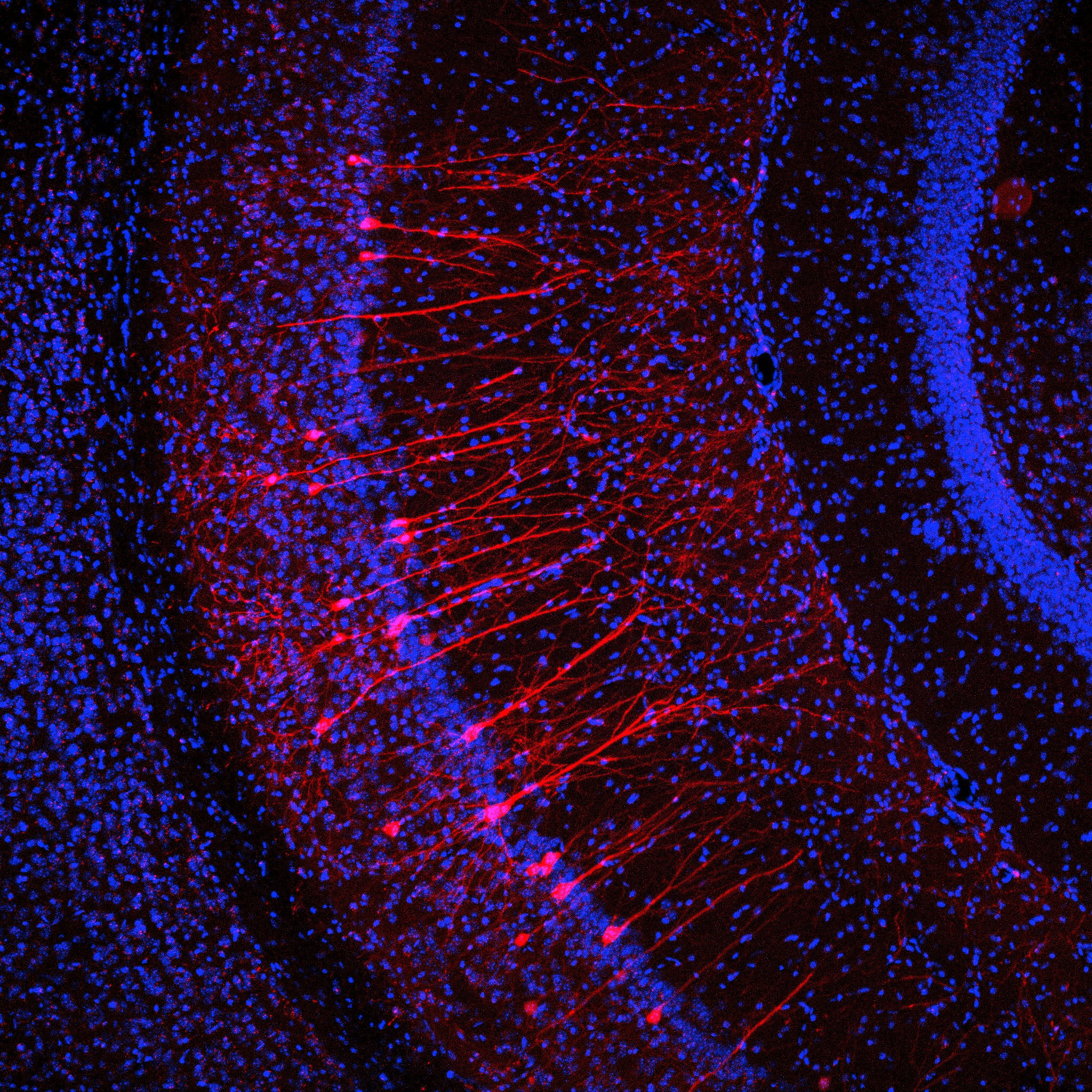
Representations of stimulus meaning in the hippocampus
Biane JS , Ladow MA , Austin Fan A , Choi HS , Zhou LZ , Hassan S , Apodaca-Montano DL, Kwon AO , Bratsch-Prince JX , and Kheirbek MA (2024)
BioRxiv
The ability to discriminate and categorize the meaning of environmental stimuli and respond accordingly is essential for survival. The ventral hippocampus (vHPC) controls emotional and motivated behaviors in response to environmental cues and is hypothesized to do so in part by deciphering the positive or negative quality of these cues. Yet, what features of the environment are represented in the activity patterns of vCA1 neurons, and whether the positive or negative meaning of a stimulus is present at this stage, remains unclear. Here, using 2-photon calcium imaging across six different experimental paradigms, we consistently found that vCA1 ensembles encode the identity, sensory features, and intensity of learned and innately salient stimuli, but not their overall valence. These results offer a reappraisal of vCA1 function, wherein information corresponding to individual stimulus features and their behavioral saliency predominates, while valence-related information is attached elsewhere.

Identifying dysfunctional cell types and circuits in animal models for psychiatric disorders with calcium imaging
Gergues MM, Lalani LK, Kheirbek MA (2024)
Neuropsychopharmacology
A central goal of neuroscience is to understand how the brain transforms external stimuli and internal bodily signals into patterns of activity that underlie cognition, emotional states, and behavior. Understanding how these patterns of activity may be disrupted in mental illness is crucial for developing novel therapeutics. It is well appreciated that psychiatric disorders are complex, circuit-based disorders that arise from dysfunctional activity patterns generated in discrete cell types and their connections. Recent advances in large-scale, cell-type specific calcium imaging approaches have shed new light on the cellular, circuit, and network-level dysfunction in animal models for psychiatric disorders. Here, we highlight a series of recent findings over the last ~10 years from in vivo calcium imaging studies that show how aberrant patterns of activity in discrete cell types and circuits may underlie behavioral deficits in animal models for several psychiatric disorders, including depression, anxiety, autism spectrum disorders, and schizophrenia. by elucidating cell types and their activity patterns. These advances in calcium imaging in pre-clinical models demonstrate the power of cell-type-specific imaging tools in understanding the underlying dysfunction in cell types, activity patterns, and neural circuits that may contribute to disease and provide new blueprints for developing more targeted therapeutics and treatment strategies.

From bile acids to melancholia
Klein AS, Kheirbek MA (2024)
Neuron
Preview for Li, Zhang et al
In this issue of Neuron, Li, Zhang, et al. find that the bile acid receptor TGR5 in the lateral hypothalamus influences neuronal dynamics underlying stress-induced depression-like behaviors. Inhibition of these neurons produces antidepressant-like effects through a circuit that includes hippocampal CA3 and dorsolateral septum, revealing a novel potential therapeutic for depression.

Neural dynamics underlying associative learning in the dorsal and ventral hippocampus
Biane JS, Ladow MA, Stefanini F, Boddu SP, Fan A, Hassan S, Dundar N, Apodaca-Montano DL, Woods NI, Kheirbek MA (2023)
Nature Neuroscience
Animals associate cues with outcomes and update these associations as new information is presented. This requires the hippocampus, yet how hippocampal neurons track changes in cue–outcome associations remains unclear. Using two-photon calcium imaging, we tracked the same dCA1 and vCA1 neurons across days to determine how responses evolve across phases of odor–outcome learning. Initially, odors elicited robust responses in dCA1, whereas, in vCA1, odor responses primarily emerged after learning and embedded information about the paired outcome. Population activity in both regions rapidly reorganized with learning and then stabilized, storing learned odor representations for days, even after extinction or pairing with a different outcome. Additionally, we found stable, robust signals across CA1 when mice anticipated outcomes under behavioral control but not when mice anticipated an inescapable aversive outcome. These results show how the hippocampus encodes, stores and updates learned associations and illuminates the unique contributions of dorsal and ventral hippocampus.

Linking external stimuli with internal drives: A role for the ventral hippocampus
Turner VS, O’Sullivan RO, Kheirbek MA. (2022)
Current Opinion in Neurobiology
The ventral hippocampus (vHPC) has long been thought of as the “emotional” hippocampus. Over the past several years, the complexity of vHPC has come to light, highlighting the diversity of cell types, inputs, and outputs that coordinate a constellation of positively and negatively motivated behaviors. Here, we review recent work on how vCA1 contributes to a network that associates external stimuli with internal motivational drive states to promote the selection of adaptive behavioral responses. We propose a model of vHPC function that emphasizes its role in the integration and transformation of internal and external cues to guide behavioral selection when faced with multiple potential outcomes.